
Guiding the scan: how AI could make portable ultrasound more accessible


A portable ultrasound device can bring imaging closer to patients, but obtaining a useful image still takes skill. At Bern University of Applied Sciences (BFH), we explore how artificial intelligence (AI) could guide non-specialists during scanning, and what must be established before that promise can translate into better access to care.
A device is only the beginning
Imagine a healthcare worker holding a portable ultrasound probe while an image appears on a tablet. The equipment is available, yet a practical question remains: where should the probe move to obtain the required view? This scene captures the challenge at the heart of our research: making imaging equipment easier to carry does not, by itself, make it easier to use.
Ultrasound acquisition depends on the operator’s skill in positioning and orienting the probe. A visible image is only a starting point: obtaining a useful view requires knowing what to look for and how to find it. This matters most where specialist expertise is hard to access.
Together with an external partner, we investigate how AI-based assistance during scanning could support healthcare workers who lack specialist ultrasound training. Our work is exploratory, and this article describes the questions that motivate it.
From an image to an actionable suggestion
The approach we envisage is a feedback loop (Figure 2). Software would assess the incoming ultrasound image, consider how well the relevant anatomy is visible and suggest an adjustment. The healthcare worker would act on that suggestion, producing a new image to assess. Related ideas have been investigated clinically in defined tasks: In a cardiac study, nurses without previous echocardiography experience acquired images of diagnostic quality for several predefined assessments after training with AI guidance [5], and a separate lung ultrasound study found that healthcare professionals using AI guidance could obtain images meeting predefined diagnostic-quality criteria [1]. These are application-specific results; they do not show that one system works across organs.
A prompt such as “rotate the probe clockwise” makes the idea tangible: it translates image analysis into something the operator can do. Delivering such a prompt reliably, however, requires more than detecting an unclear image. The system must also judge whether a particular adjustment is likely to help. The example illustrates a proposed interaction, not an existing capability.
Three elements underpin our ambition: assessing images, giving understandable guidance and processing information efficiently enough for portable use. Feedback must arrive quickly enough to remain relevant as the operator moves the probe, so balancing speed, image assessment and the limited computing resources of a portable device is part of the research problem.
We focus on acquiring the image; interpreting it to reach a diagnosis is a separate task.
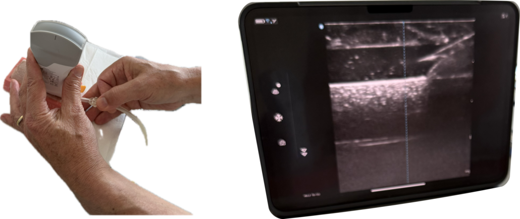
Figure 1: Portable ultrasound imaging setup: handheld transducer (left) and tablet-based B-mode display (right).

Figure 2: Conceptual guidance loop. Software would assess an image and suggest an adjustment; the healthcare worker would act on that suggestion and acquire a new image. The figure describes a research aim, not a validated system architecture.
What would make guidance useful in practice?
Guidance can only be as good as the system’s understanding of what the probe is showing. Ultrasound is a demanding setting for computer vision: images are noisy, brightness varies across the field of view, and the appearance of the same anatomy changes with the probe’s position and the patient. Earlier ultrasound research already combined local and global image statistics in an adaptive segmentation framework designed to handle noise and spatial intensity inhomogeneity [2]. Before software can advise on where to move, it must recognise which structures are visible and which are missing.
This is why we treat image understanding as a foundation for guidance. Segmentation, which separates an image into meaningful regions, and a broader understanding of anatomy would give a guidance system something concrete to reason about: what is in view, how clearly, and what a better view would contain. Our current work therefore studies the output of portable devices and how it can be processed, addressing individual components before attempting to connect them into a complete system.
Recent research on ultrasound foundation models suggests a route towards such understanding. These models learn reusable features from large image collections and can then be adapted to different organs and tasks. For example, USFM was self-supervised pre-trained on a multi-organ ultrasound database containing more than two million images and achieved strong performance in segmentation and classification compared with representative ultrasound image-analysis models in the reported experiments [4]. Classification assigns images to categories. Reusing a shared model could reduce the labelled data needed for each application. Whether such a model suits a particular device, clinical task and live guidance workflow would still need evaluation.
Understanding an image is not the same as knowing how to improve it. A method might recognise anatomy consistently and still provide too little information to choose a helpful adjustment. Evaluating image analysis therefore answers a different question from evaluating guidance for operators. Connecting the two requires a clear account of what information guidance needs and how image analysis can supply it.
How could we tell whether it helps?
Clinical specialists, technical researchers and the device partner each contribute to answering three questions. Technical performance alone does not describe how a system works in practice. The DECIDE-AI reporting guideline for early clinical evaluation of AI-based decision support systems similarly emphasizes describing not only the AI system itself, but also its interaction with users and the clinical environment in which it is deployed [7].
Does the advice help users obtain the intended image? A meaningful assessment would begin with a defined clinical task and criteria for judging image usefulness, which clinical specialists would need to help establish. Comparing acquisition with and without guidance, under appropriate conditions, could then test whether assistance makes a difference. The data collection and evaluation design still need to be specified; these are questions for future work, not results of a completed study.
Can users understand the advice and recognise its limits? A technically plausible suggestion is useful only if the operator can act on it. Better images and faster scanning are also separate outcomes: A laboratory study of novice operators performing a focused abdominal ultrasound view found higher diagnostic image quality with AI guidance, although examination times were initially longer [3]. Evaluation should examine how people interpret instructions, what training they need and what happens when the system cannot offer reliable guidance. Assistance should support professional judgement and make its limits clear. The ambition to help non-specialists does not mean that training can be dispensed with.
Can the approach work in the intended setting? Portable processing is one requirement; the surrounding workflow matters too. We need to address how images are collected and handled, how assistance fits into clinical practice and when specialist support is needed.
From wider availability to meaningful access
For the healthcare worker in the opening scenario, the potential value is concrete: help at the moment of uncertainty about how to obtain a useful view. If such support proves effective, it could make portable imaging more usable where expertise and resources are limited. Research in Zambia explored a different route to lowering expertise barriers: estimating gestational age from blind ultrasound sweeps acquired by untrained nurse midwives using low-cost, battery-powered ultrasound equipment [6]. That work interprets recorded sweeps rather than instructing probe movement. Guidance might also support learning, although that benefit would need its own evaluation.
This is our societal motivation. Better access to care and reduced inequalities are intended contributions, not outcomes established by our exploratory work. Their realisation would depend not only on technical capability but also on how a working system reaches people and supports their practice.
A focused clinical application would make that ambition testable. Instead of judging success against a broad promise of assistance for many organs, we can ask whether guidance helps a particular group of users with a clearly defined task.
Access must include usable support
Portable ultrasound and AI guidance could complement each other: one brings equipment closer to where it is needed, while the other aims to help people obtain useful images. Our research addresses the gap between that promise and a dependable tool. Its wider lesson for digital transformation is that access involves more than possessing technology: it also requires support that people can understand and use effectively, together with evidence of where that support helps and where its limits lie.
References
[1] Cristiana Baloescu, John Bailitz, Baljash Cheema, et al. Artificial intelligence–guided lung ultrasound by nonexperts. JAMA Cardiology, 10(3):245–253, 2025.
[2] Djamal Boukerroui, Atilla Baskurt, J. Alison Noble, and Olivier Basset. Segmentation of ultrasound images—multiresolution 2D and 3D algorithm based on global and local statistics. Pattern Recognition Letters, 24(4–5):779–790, 2003.
[3] I-Min Chiu, Chun-Hsiang Ryan Lin, Fu-Fan Frank Yau, et al. Use of a deep-learning algorithm to guide novices in performing focused assessment with sonography in trauma. JAMA Network Open, 6(3):e235102, 2023.
[4] Jing Jiao, Jin Zhou, Xiaokang Li, Menghua Xia, Yi Huang, Lihong Huang, Na Wang, Xiaofan Zhang, Shichong Zhou, Yuanyuan Wang, and Yi Guo. USFM: A universal ultrasound foundation model generalized to tasks and organs towards label efficient image analysis. Medical Image Analysis, 96:103202, 2024. https://doi.org/10.1016/j.media.2024.103202.
[5] Akhil Narang, Richard Bae, Hyung Hong, et al. Utility of a deep-learning algorithm to guide novices to acquire echocardiograms for limited diagnostic use. JAMA Cardiology, 6(6):624–632, 2021.
[6] Teeranan Pokaprakarn, Juan C. Prieto, Joan T. Price, et al. AI estimation of gestational age from blind ultrasound sweeps in low-resource settings. NEJM Evidence, 1(5):EVIDoa2100058, 2022.
[7] Baptiste Vasey, Myura Nagendran, Bruce Campbell, et al. Reporting guideline for the early-stage clinical evaluation of decision support systems driven by artificial intelligence: DECIDE-AI. Nature Medicine, 28:924–933, 2022.

Dr Albin Grataloup is a postdoctoral researcher in data science at the Bern University of Applied Sciences. His research focuses on personalized and privacy-preserving learning methods applied, among others, to medical and well-being monitoring, generative and recommendations systems.
 Christoph Paulus is a professor of computer science at Bern University of Applied Sciences and heads the Computer Vision Laboratory – which also focuses on simulations and artificial intelligence.
 Contributions as RSS
 Comments as RSS

Leave a Reply
Want to join the discussion?Feel free to contribute!